

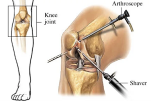
Knee arthroscopy is one of the most frequently used procedures for the diagnosis and treatment of knee injuries. This minor surgical procedure is done using an instrument called an arthroscope. While the knee is the joint most often viewed and operated using the arthroscope, other joints such as the shoulder, elbow, ankle, hip and wrist can also be viewed using this instrument.
What are the causes of the knee pain?
Injuries, overuse, weakness or aging can harm your knees and cause pain and discomfort. What are the benefits of knee arthroscopy? Knee arthroscopy is an effective tool in diagnosing your joint condition and for confirming treatment for knee problems such as meniscus tears and cartilage wear. An arthroscopy can ultimately provide relief from knee pain and improve mobility. Maintaining a normal and active lifestyle with greater comfort is a key benefit of this procedure.
What are the risks of knee arthroscopy?
While knee arthroscopy is a safe procedure for the treatment of knee injuries, there are some risks that patients should be aware of. Depending on the patient and joint condition, these risks may include swelling and stiffness of the joint, bleeding, blood clots, infection, or continuing knee problems.
How long is the recovery period after knee arthroscopy?
The recovery period after knee arthroscopy surgery depends on the patient and his or her condition. For most cases, patients can return to office work within a week. And many return to a more active and normal lifestyle within one to two months.
Bilateral arthroscopy is a surgical procedure performed on both joints of a particular body part simultaneously, typically referring to the knees or shoulders.
In the case of bilateral arthroscopy, the procedure involves examining and addressing issues in both joints during a single surgical session. This approach offers several advantages, including reduced overall surgical time, decreased anesthesia exposure, and potentially shorter recovery period compared to separate surgeries on each joint.
Following both knee surgery, the patient typically undergoes a structured rehabilitation program to promote healing, restore joint function, and optimize recovery. This program may include physical therapy, pain management, and exercises to strengthen the surrounding muscles and improve range of motion.
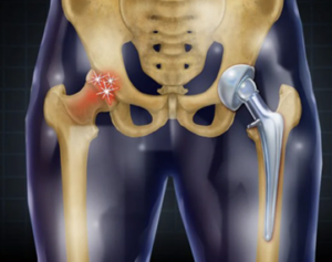
Total hip replacement (or hip arthroplasty) is a technique that has become widespread in recent years in response to the need for improving hip joints that have been damaged by injury or arthritis. Joint replacement surgery may offer the best treatment option for long-term improvement for the hip joint when other treatments have proven inadequate. In most cases, having a total hip replacement reduces joint pain and means a return to pain-free movement.
What causes hip pain?
The human hip is a ball and socket joint. It is the most flexible and free-moving joint in the body, and can move backwards and forwards, to the side, and can perform twisting motions. Full function of the hip is dependent on the coordination of bones, muscles, tendons, ligaments and nerves.
Hip pain can be caused by a variety of factors, including:
•An injury that does not heal properly.
•A chronic illness.
•Normal wear and tear from years of constant use.
•Severe arthritic conditions, especially osteoarthritis.
•Injuries as a result of trauma, such as a hip fracture or dislocation caused by a fall.
If hip replacement surgery is the best treatment option for you, your physician will refer you to an orthopedic surgeon with expertise in this procedure. Our orthopedic surgeon will evaluate your hip joint with a full physical exam and X-rays and develop a treatment plan about how surgery can best benefit you. Our surgeon will also ask you about any past medical problems.
What are the benefits of hip replacement?
Relief from pain is the greatest benefit and the major reason for hip replacement surgery. The procedure offers other benefits, such as:
Improved movement, strength and coordination of the torso and leg. The ability to walk, climb stairs and maintain an active lifestyle in greater comfort.
What are the risks of surgery?
There are possible risks and complications that may happen through hip replacement surgery associated with anesthesia, including respiratory or cardiac malfunction. Other complications include:
•Blood clots
•Infection Injury to nerves and blood vessels
•Fracture
•Weakness
•Stiffness or instability of the joint
•Need for additional surgeries
•Dislocation
also known as ACL (anterior cruciate ligament) surgery, is a procedure performed to reconstruct or repair a torn ACL in the knee joint. The ACL is a cruciate ligament that provides stability to the knee and helps prevent excessive forward movement of the shin bone.
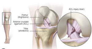
During the surgery, the torn ACL is either repaired by reconnecting the torn ends or reconstructed using a graft, typically taken from the patient’s own hamstring tendon or patellar tendon, or a donor graft. Our surgeon uses arthroscopic techniques, where small incisions and an arthroscope are used to visualize and access the knee joint.
After cruciate ligament surgery, the recovery procedure typically involves the following:
•Immobilization: Initially, the knee may be placed in a brace or immobilizer to protect the surgical site and aid in healing.
•Physical therapy: Physical therapy plays a crucial role in the recovery process. It usually begins shortly after surgery and focuses on reducing pain and swelling, regaining range of motion, and strengthening the surrounding muscles.
•Weight-bearing and walking: The ability to bear weight on the operated leg and gradually resume walking is a significant milestone in the recovery process. The timing and extent of weight-bearing depend on the surgeon’s instructions and the individual’s progress.
•Rehabilitation exercises: Specific exercises are prescribed to gradually increase strength, stability, and flexibility of the knee. These exercises may include leg presses, hamstring curls, squats, balance exercises, and agility drills.
•Return to sports and activities: The timing for resuming sports or high-impact activities varies depending on individual factors, such as the healing progress, strength, and stability of the knee. The surgeon or physical therapist will guide the patient on gradually returning to these activities and may recommend the use of knee braces for additional support.
It’s important to note that the recovery period can vary for each individual, typically ranging from several months to a year, depending on the extent of the injury, surgical technique, and commitment to rehabilitation. Close adherence to the post-operative instructions and regular follow-up appointments with the healthcare team are essential to achieve a successful recovery and regain full function of the knee.
Interventional knee arthroscopy is a minimally invasive surgical procedure performed to diagnose and treat various knee joint conditions using specialized instruments and an arthroscope. It involves making small incisions in the knee to access and visualize the joint.
During the surgery, our surgeon can perform a variety of interventions, including removing loose bodies, repairing torn cartilage or ligaments, debriding damaged tissue, and addressing joint inflammation. The arthroscope provides a clear view of the joint, allowing for precise and targeted treatments.
The recovery period following interventional knee arthroscopy can vary depending on the specific procedure performed and the individual’s healing capacity. However, in general, the following elements are typically involved in the recovery process:
•Immobilization: Initially, the knee may be immobilized with a brace or splint to protect the joint and facilitate healing.
•Pain management: Medications and techniques for managing post-operative pain and swelling are commonly employed, as prescribed by the surgeon.
•Rehabilitation exercises: Physical therapy plays a crucial role in the recovery process. It typically begins shortly after surgery and focuses on restoring range of motion, strengthening the knee muscles, and improving overall joint function.
•Weight-bearing and walking: The ability to bear weight on the operated leg and gradually resume walking is an important aspect of recovery. The timing and extent of weight-bearing depend on the specific procedure and the surgeon’s instructions.
•Return to normal activities: The timeline for returning to normal activities can vary depending on the extent of the procedure and the individual’s progress. The surgeon or physical therapist will provide guidance on when it is safe to resume daily activities and potentially return to sports or higher impact exercises.
The recovery period for interventional knee arthroscopy is typically shorter compared to more extensive knee surgeries. However, it can still take several weeks to a few months to fully recover and regain optimal knee function. Compliance with post-operative instructions, regular physical therapy sessions, and follow-up appointments with the healthcare team are crucial for a successful recovery.
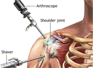
Shoulder impingement syndrome – characterized by bursitis, inflammation, sometimes tearing of the rotator cuff, and bone spur formation in the shoulder — produces pain and limits range of motion.
Symptoms include discomfort and, sometimes, acute shoulder pain when raising the arms above the head, reaching into a back pocket or while sleeping on the affected shoulder. People who frequently work with their arms above their heads may be more likely to get shoulder impingement syndrome.
What are your treatment options?
Before beginning treatment for shoulder pain, your doctor may take an X-ray. You doctor will also take a thorough history and examine you to ensure that the pain is not due to a problem in another area of the body.
Medical Process
Non-operative measures, including activity modification, physical therapy, anti-inflammatory medication, rehabilitation and, if appropriate, cortisone injection, are effective for more than 90 percent of patients in resolving the shoulder instability symptoms within three to six months of onset.
Surgical Process
Our doctor may suggest surgery for the patient who has not benefited from the non-operative management. The surgery may be performed arthroscopically or open, depending on which method the surgeon feels is more appropriate. This procedure improves function in about 85 percent of patients. The majority of cases are performed arthroscopically at our Hospitals and Clinics.
Arthroscopic subacromial decompression requires at least two 5 mm incisions. The arthroscope is introduced through the skin and deltoid muscle to enter the shoulder joint. Once in the joint, a methodical inspection of the tissues is performed looking for tears in ligaments, muscles and tendons. Loose bodies may be found and an overall assessment of the joint surface is made. With this technique, the biceps tendon, rotator cuff tendons and joint can be examined for injuries that may not be evident on an MRI scan or with open surgery. After the joint is examined, the arthroscope is withdrawn through the rotator cuff tendons and is placed in the space above the rotator cuff. It is in this space that the surgeon smoothes off the spur that is contributing to inflammation and impingement on the rotator cuff. Arthroscopic surgery allows for a shorter recovery time and predictably less pain in the first few days following the procedure than does open surgery.
Osteotomy literally means “cutting the bone”. In knee osteotomy, the tibia (shinbone) or femur (thigh bone) is cut and then reshaped to relieve pressure on the knee joint.
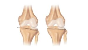
Knee osteotomy is used when the patient has early-stage osteoarthritis that damages only one side of the knee joint. By displacing the weight of the damaged side of the joint, an osteotomy can relieve pain and significantly improve function in an arthritic knee.
Explanation
Osteoarthritis can develop when the bones of your knee and leg are not aligned properly. This can put extra stress on the inner (medial) or outer (lateral) side of your knee. Over time, this extra pressure can wear away the soft articular cartilage that protects the bones and causes pain and stiffness in the joint.
Advantages and disadvantages
There are three goals in knee osteotomy:
• Transferring from the arthritic part of the knee to a healthier area
• To correct poor knee alignment
• To prolong the life of the knee joint
One advantage of the procedure is that by preserving your own knee anatomy, a successful osteotomy can delay the need for joint replacement for several years. Another advantage is that there are no restrictions on physical activities after an osteotomy. You can participate in your favorite activities, even high-impact exercise.
Osteotomy has its disadvantages. For example, pain relief after osteotomy is not predictable compared to partial or total knee replacement. Recovery from an osteotomy is typically longer and more difficult because you may not be able to immediately lose weight on your operated knee. In some cases, having an osteotomy can make knee replacement surgery more difficult later on. Knee osteotomy has become less common as the results achieved with total knee replacement and partial knee replacement have been so successful. Still, it remains an option for many patients. Most osteotomies for knee arthritis are done on the tibia (shinbone) to correct a springy alignment that puts too much pressure on the inside of the knee.
During this procedure, a wedge of bone is removed from the outside of the tibia, on the healthy side of the knee. When the surgeon closes the wedge, he straightens the leg. This brings the bones closer to the healthy side of the knees and creates more space between the bones on the damaged, arthritic side. As a result, the knee can move more evenly, relieving pressure on the painful side. This procedure is sometimes called a “high tibial osteotomy.” Osteotomies of the thigh bone (femur) are performed using the same technique.
What is spine surgery?
The spinal cord provides communication between the brain and other organs with the help of nerves and is located in a thick membrane just like the brain. It extends from the skull to the waist in a bone called the vertebra. While transmitting the stimuli from the brain to the arms and legs or other organs, data from different organs are transmitted to the brain in this way. In case of any compression that may occur on the spinal cord due to these functions, patients can become paralyzed. In the treatment of these diseases, the “Microsurgery” method is the most ideal operation technique.
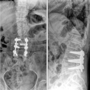
With the microsurgery technique, complex (heavier-difficult) spine surgeries can be successfully performed in the hybrid operating room. In which diseases is spine surgery used in the treatment?
What are spinal diseases?
Spinal diseases can be divided into 4 groups.
• Congenital spinal diseases (such as spina bifida waist opening, cerebellum prolapse, tense spinal cord, scoliosis)
• Degenerative (hernia, narrow canal, scoliosis, spinal slippage)
• Those due to trauma (Fracture, spinal cord injuries)
• Spine and spinal cord tumors
Spine health is teamwork
The spine, which is formed by the union of 33 vertebral bones, is the most important indicator of health both because of the structures it contains and the structures it supports. At Sanita Group Hospitals and Spine Surgery Clinic, spinal health is approached from a 360-degree perspective. A holistic perspective is adopted in the our Spine Surgery Clinic, which consists of spine surgeons, algologists, physical therapists and psychiatrists. Personalized treatment options are offered with a multidisciplinary approach.
The most common spinal diseases
• Degenerative spine diseases such as low back or neck hernia
• Spinal curvatures such as scoliosis or kyphosis
• Traumas of the spine area
• Spinal tumors
• Spine infections When should you consult a doctor?
• Low back pain and pain radiating to the leg
• Neck pain and pain radiating to the arms and back
• Numbness and weakness in the legs
• Numbness and weakness in the hands
• Shoulder imbalance
• Breast asymmetry
• Posture disorder
Spine surgery, also known as 7-Up surgery, refers to a specific type of surgical intervention aimed at treating spinal disorders and deformities, such as scoliosis. The name “7-Up” is derived from the seven vertebrae that are typically fused together during the procedure.
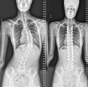
During 7-Up surgery, our surgeon makes incisions in the back and utilizes screws, rods, and bone grafts to stabilize and realign the spine. The procedure involves straightening the spinal curvature and fusing the affected vertebrae to maintain proper alignment. This helps alleviate pain, improve spinal stability, and prevent further progression of the spinal deformity.
The recovery duration following 7-Up surgery can vary depending on the specific details of the procedure, the individual’s overall health, and the extent of spinal correction required. Generally, the following elements are involved in the recovery process:
Hospital stay: After surgery, the patient typically stays in the hospital for a few days to a week for close monitoring of post-operative care, pain management, and physical therapy initiation.
Pain management: Medications are prescribed to manage post-operative pain and discomfort.
Physical therapy: Physical therapy is an integral part of the recovery process and aims to restore strength, flexibility and function of the spine. Our therapists guide the patient through specific exercises and techniques to promote healing and improve mobility.
Restricted activities: Certain activities, such as heavy lifting and strenuous exercises, may be restricted for a period of time to allow the spine to heal properly.
Gradual return to normal activities: The timing for returning to normal activities varies depending on the individual’s healing progress and the surgeon’s guidance. It may take several months before the patient can resume regular daily activities.
The complete recovery duration for 7-Up surgery can extend from several months to a year, and sometimes longer, as it involves the fusion of multiple vertebrae. It is crucial to follow our surgeon’s instructions regarding post-operative care, physical therapy, and regular check-ups to ensure a successful recovery and optimize the long-term outcome of the procedure.
It’s important to note that the information provided here is a general overview, and the specific treatment, surgical techniques, and recovery duration may vary depending on your health needs and the recommendations of our specialist healthcare team.


